Limpets graze on Benthic microalgae. These form films on mud surfaces and hard rocky substrates and include diatoms, cyanobacteria and microscopic sporeling stages of macroalgae. These organisms form a thin biofilm layer on the substratum surface.
To help limpets scrape the microalgae off of the rock limpets like other gastropods have a radula. A typical radula comprises a number of bilaterally-symmetrical rows of teeth rooted in a radular membrane. Some species have teeth that bend with the membrane as it moves over the odontophore, whereas in other species, the teeth are firmly rooted in place, and the entire radular structure moves as one entity.
Radular membrane is The elastic, delicate radular membrane may be a single tongue, or may split into two (bipartite).
Odontophore
The odontophore is the tongue of flesh underlying the radular membrane, and controls the organ's protrusion and return. It can be likened to a pulley wheel over which the radular 'string' is pulled.


 The mantle (the tan ring around the visceral mass) is then peeled back to reveal the head and one tentacle either side.


To help limpets scrape the microalgae off of the rock limpets like other gastropods have a radula.
 A typical radula comprises a number of bilaterally-symmetrical rows of teeth rooted in a radular membrane. Some species have teeth that bend with the membrane as it moves over the odontophore, whereas in other species, the teeth are firmly rooted in place, and the entire radular structure moves as one entity.
Radular membrane is The elastic, delicate radular membrane may be a single tongue, or may split into two (bipartite).
Odontophore
The odontophore is the tongue of flesh underlying the radular membrane, and controls the organ's protrusion and return. It can be likened to a pulley wheel over which the radular 'string' is pulled.
 |
| Radula Mechanism |

To observe a limpet radula the visceral mass of the limpet needs to be removed from the shell this is easily done by boiling the limpet in its shell.


The body of the limpet will then easily fall out of the shell. The next step is to pin the limpet down to stop it sliding around.

 The mantle (the tan ring around the visceral mass) is then peeled back to reveal the head and one tentacle either side.
A small incision is then made between the eyes and the radula can easily be pulled out of the odontophore.

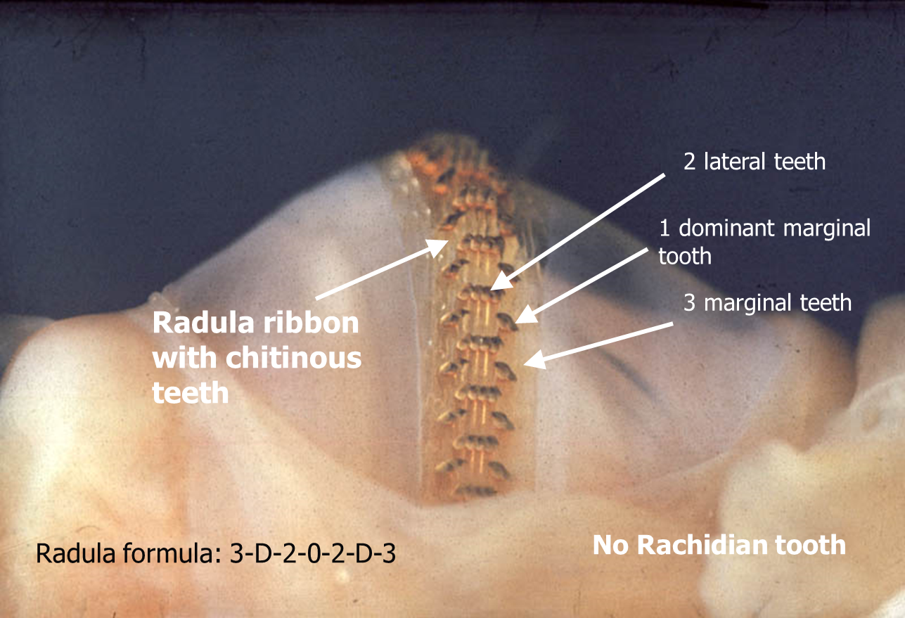
This is the radula from a 3.1 cm limpet the radula measured 6.4 cm this is because the teeth are worn out and are constantly replaced. The thicker end on the right of the photo is where it attaches to the odontophore.


These images are taken down the eyepiece of a compound microscope you can clearly see the two sets of 1 dominant marginal tooth and the 2 lateral teeth . You can just see the feathery edge suggesting the 3 marginal.
No comments:
Post a Comment